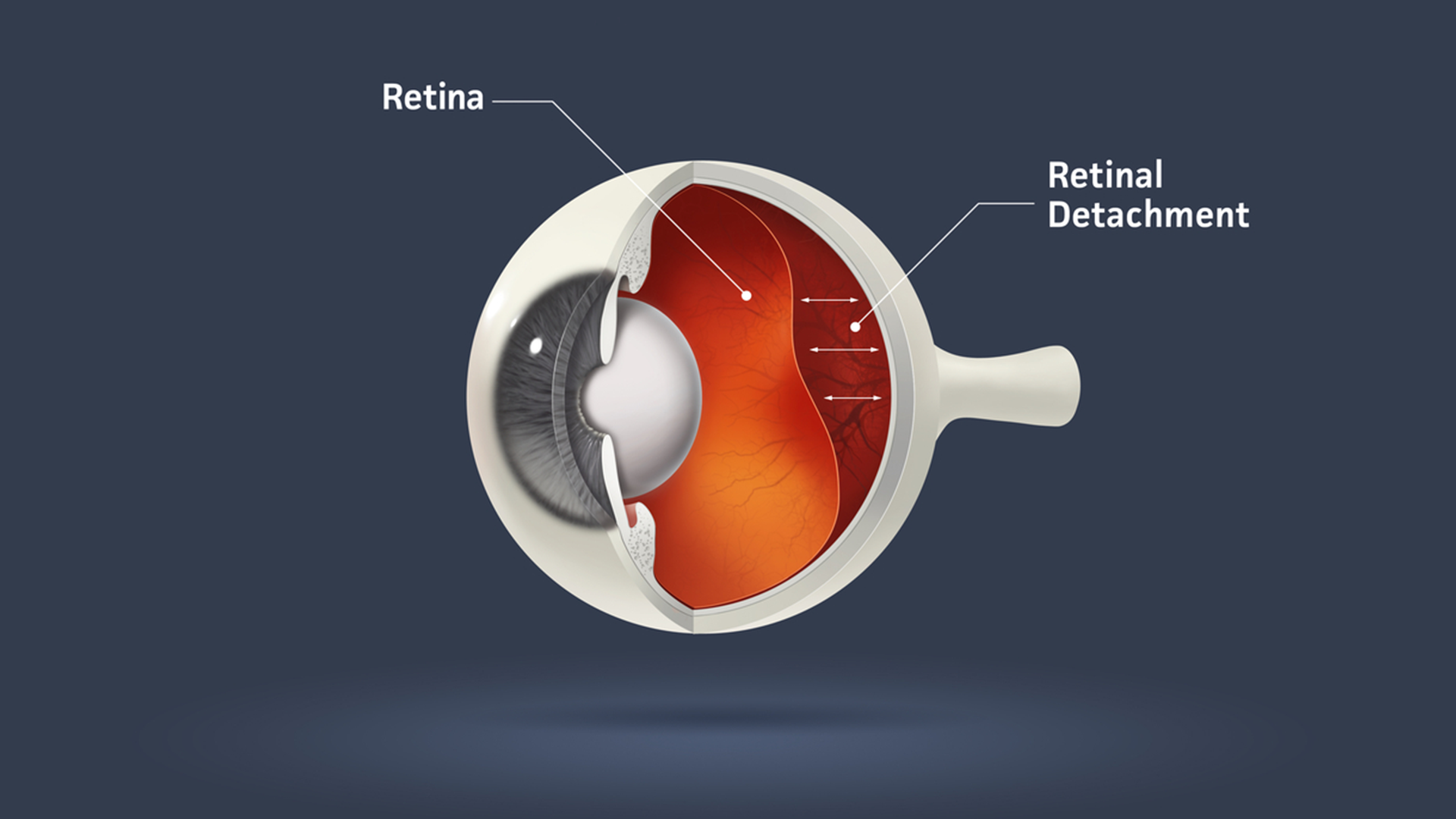
Retinal Detachment
Retinal detachment is separation of the retina from the underlying layers that line the inner wall of the eye. Through the retinal tear, liquid from the vitreous may pass through the tear, and detach the retina. As the fluid accumulates, more and more of the retina detach. Detached retina loses its function; hence the person with retinal detachment loses vision suddenly or gradually.
Although anyone can develop a retinal detachment, some people are at a high risk. Myopic patients (nearsighted people), those who have ‘weak areas’ in the retina, known as lattice degeneration, those who have had significant eye injuries, and those with a family history of retinal detachment are at higher risk of retinal detachment. Retinal detachment can also occur following cataract surgery. Retinal Detachment is an emergency – the earlier the treatment, the better the vision.
CAUSES
The vitreous-a gel like material is present is maximum areas of eye. Meanwhile, the presences of retinal tear allow gel from vitreous space to pass through the hole and flow between the retina and the back wall of the eye. As a result the retina detaches from its underlying layer of support tissue at the back of the eye. The detached area of the retina will not function properly and if not treated initially the whole retina will peel off and the person may lose his/her vision.
SYMPTOMS
Most people notice floaters and flashes before the retina detaches. As the detachment increases a gradually enlarging dark shadow engulfs vision. It may appear as a curtain or a shade drawn slowly across the field of vision. Central retina is the area that helps one see fine detail also allowing one to read small print. When retinal detachment progresses to involve the central retina, the reading ability is lost. In complete retinal detachment, one may just see light and no other details.
